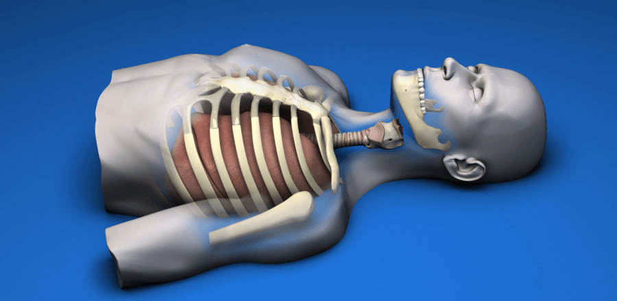
SAM 5: Emergency Surgical Airway Model
SAM 6: Pleural Space Decompression Model
SAM 9: Chest Wall Fixation Model
The lower jaw, anatomical landmarks of the throat and root of neck are reproduced to allow needle cricothyroidotomy, tube insertion and tracheostomy to be executed. This model can be combined with SAM 3.1 Proximal Humerus Intraosseous Infusion and SAM 6 Pleural Space Decompression Models as illustrated. Chosen patterns of facial trauma can be represented to indicate upper airway obstruction. After successful intubation, the model’s chest will rise and fall upon positive pressure ventilation.
The laryngeal and crycoid cartilages along with the adjoining section of the trachea can be exchanged through the back of the model for repeated use and the overlying skin repaired.
A smaller version restricted to the anatomy of the neck can also be produced. Pleural space decompression is required in the management of tension and simple pneumothorax, and haemothorax. Needle thoracocentesis is a recognised method for equilibrating intra-pleural pressure with atmospheric pressure and so relieving tension pneumothorax.
A cannula of suitable length is inserted into the 2nd intercostal space in the mid-clavicular line. Finger thoracostomy is increasingly recognised as a more reliable method of decompression the pleural space. A 2.5cm incision is made along the upper border of the 5th rib in the anterior axillary line. Blunt dissection over the superior border of the rib enables entry to the pleural space. A single figure is introduced into the pleural space to ensure decompression. Intercostal drain insertion is then accomplished through the finger thoracostomy wound.
Anterior rib fracture fixation can be executed on this model, of a simplified version will be available for training solely in this technique.


